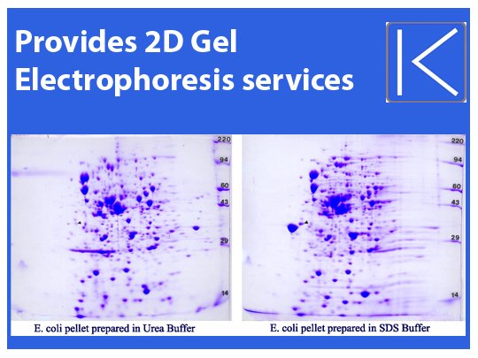
2D Protein Gel Analysis is a widely used technique in the field of proteomics that allows for the simultaneous separation and analysis of complex protein mixtures. This powerful method provides valuable insights into protein expression, post-translational modifications, and protein-protein interactions. In this article, we will explore the essential steps involved in 2D Protein Gel Analysis, focusing on the techniques and methodologies employed for sample preparation, electrophoresis, protein staining and visualization, image analysis, protein identification, quality control, and troubleshooting. Additionally, we will discuss recent advancements in the field and the future directions that are shaping the way we study and understand proteins. Whether you are a researcher new to 2D Protein Gel Analysis or seeking to enhance your knowledge, this article, presented by Kendrick labs, will serve as a comprehensive guide to the fundamental steps and advancements in this essential analytical tool.
Introduction to 2D Protein Gel Analysis
Overview of 2D Protein Gel Analysis
Picture this: you’re a detective, but instead of looking for clues to solve a crime, you’re on the hunt for proteins. Welcome to the fascinating world of 2D protein gel analysis. This technique allows scientists to separate and analyze thousands of proteins from a single sample, giving us valuable insights into their functions and interactions.
In a nutshell, 2D protein gel analysis involves two steps: separating proteins based on their charge and size using a clever combination of electrophoresis techniques, and then visualizing them using various staining methods. By analyzing the resulting gel images, scientists can identify and compare proteins across different samples, helping us understand diseases, discover potential drug targets, and uncover the mysteries of molecular biology.
Importance and Applications of 2D Protein Gel Analysis
Wondering why 2D protein gel analysis is such a big deal? Well, proteins are the workhorses of living organisms, carrying out vital functions like catalyzing reactions, transporting molecules, and providing structural support. Understanding the complex world of proteins is key to unlocking the secrets of life itself.
2D protein gel analysis plays a crucial role in many areas of research, including molecular biology, medicine, and biotechnology. It helps us study protein expression patterns in different tissues, identify biomarkers for diseases, investigate protein-protein interactions, and even discover new proteins. So whether you’re a scientist searching for answers or just a curious soul fascinated by the wonders of biology, 2D protein gel analysis is your ticket to unraveling the mysteries of the protein universe.
Sample Preparation for 2D Protein Gel Analysis
Protein Extraction and Lysis
Before you can embark on your protein separation adventure, you need to extract those pesky proteins from your sample. This often involves breaking open cells and releasing the proteins using techniques like sonication, grinding, or chemical lysis. It’s like setting the proteins free from their cellular prison!
Protein Quantification and Normalization
Now that your proteins are liberated, it’s time to measure and compare their quantities across different samples. Protein quantification methods, such as the Bradford or BCA assay, help you determine the concentrations of your protein extracts. This step is important for ensuring equal loading of proteins onto the gels and producing reliable results. Think of it as making sure all your protein guests are on an equal playing field.
Sample Preparation Techniques
To prepare your protein extracts for separation, you’ll need to do a bit of magic (not the Hogwarts kind, unfortunately). Sample preparation techniques, like reducing and alkylating agents, help ensure that your proteins are in their most suitable form for separation. By breaking disulfide bonds and preventing reformation, these techniques make sure your proteins are ready to showcase their true personalities on the gel.
Electrophoresis: Separation of Proteins
Theory and Principles of Electrophoresis
Get ready to party, because it’s time for electrophoresis! In this step, you’re going to put those proteins on the dance floor (also known as a gel) and make them move to the beat of an electric field. By applying an electrical current, proteins migrate through the gel based on their charge and size, giving you a separation that would make any DJ proud.
Equipment and Gel Types
To get this party started, you’ll need some specialized equipment and gel types. Electrophoresis chambers, power supplies, and combs are your essential tools for creating the perfect separation environment. The type of gel you choose, such as polyacrylamide or agarose, depends on the size range of proteins you’re interested in. It’s like picking the right music playlist to keep the dance floor grooving.
Gel Casting and Running Conditions
Now that you have your equipment and gel ready, it’s time to cast and run the gel. Like baking a cake, you carefully mix your gel ingredients, pour them into the casting apparatus, and let them solidify. Once the gel is ready, you load your protein samples and let the electric current do its thing. Running conditions, such as voltage and running time, affect the separation quality, so it’s important to find the sweet spot for your proteins to strut their stuff.
Protein Staining and Visualization
Staining Methods for Protein Visualization
Congratulations, you’ve successfully separated your proteins! Now it’s time to give them some color. Staining methods, like Coomassie Brilliant Blue or silver staining, help you visualize the protein bands on the gel. It’s like giving your proteins a makeover, so they can shine and catch your eye.
Image Acquisition and Documentation
Once you’ve stained your gel, you’ll want to capture an image to document your protein separation masterpiece. Imaging systems and cameras come into play here, helping you snap a photo that will immortalize your hard work. With your gel image in hand, you can analyze and compare protein expression patterns, identifying potential heroes or culprits in your biological investigations.
So there you have it, the essential steps for 2D protein gel analysis laid out for your scientific pleasure. With these techniques, you’re armed and ready to explore the intricate world of proteins and unlock the secrets they hold. Happy gel-ing!
Image Analysis for Quantification
Basics of Image Analysis
Image analysis may sound like a fancy term, but it’s actually just a way to make sense of the chaotic mess of spots on your 2D protein gel. It involves converting your gel image into digital data that you can work with. Think of it like turning your grandma’s secret recipe into a Pinterest-friendly format.
Spot Detection and Matching
Once you have your digital gel image, the next step is to identify and match the spots on the gel. It’s like playing a protein version of “Where’s Waldo?” but without the striped shirt and the beanie. Spot detection algorithms take care of the heavy lifting here, automatically finding and highlighting the spots on your gel image. It’s like having a superpowered spotter (pun intended) that never skips leg day.
Quantification and Statistical Analysis
Now that you’ve found your spots, it’s time to crunch some numbers. Quantification involves measuring the intensity or volume of each spot to determine the relative amounts of protein present. It’s like analyzing the different flavors in a box of assorted chocolates to see which one is the most popular (spoiler alert: it’s always the caramel-filled one). Statistical analysis then helps you make sense of all that data and determine if your findings are statistically significant or just a fluke. It’s like having a trusty calculator that tells you if your results are legit or if you need to go back to the lab and try again.
Protein Identification and Database Searching
Protein Digestion and Peptide Extraction
Now that you know how much protein is lurking in each spot, it’s time to play detective and figure out their identities. Protein digestion involves breaking down the proteins into smaller peptides, like dissolving a puzzle into tiny pieces. Peptide extraction then helps separate and isolate these peptides so they can be analyzed further. It’s like assembling a puzzle, but with teeny-tiny puzzle pieces and a microscope.
Mass Spectrometry Analysis
Mass spectrometry, or “mass spec” for short, is the superhero of protein identification. It helps determine the mass and charge of the peptides, giving you clues about their identities. It’s like having a molecular superpower that can tell you if a peptide is more like Batman or Superman (spoiler alert: proteins don’t wear capes). Mass spec analysis is like your lab partner who always has the answers and saves the day.
Database Searching and Protein Identification
Once you have the mass spec data, it’s time to play matchmaker with protein databases. Database searching involves comparing the mass spec data to known protein sequences to find a match. It’s like swiping through a protein Tinder, searching for the perfect match. When you find a match, you can finally unveil the identity of the mystery protein. It’s like solving a whodunit and saying, “It was Colonel Mustard, with the lead pipe, in the library!”
Quality Control and Troubleshooting
Common Issues and Challenges
No matter how skilled you are, sometimes things don’t go according to plan. Common issues and challenges can include smudged spots, uneven gel loading, or even mischievous lab gremlins. It’s like facing unexpected plot twists in a suspenseful thriller. But fear not, because every problem has a solution (except for the lab gremlins, they’re a lost cause).
Quality Control Measures
To ensure that your results are as accurate as possible, it’s important to implement quality control measures. This involves double-checking your experimental procedures, using reference standards or internal controls, and keeping detailed records of your experiments. It’s like being your own personal lab inspector, making sure everything is up to code.
Advancements and Future Directions in 2D Protein Gel Analysis
Recent Technological Advancements
The world of 2D protein gel analysis is constantly evolving like a Kardashian family drama. Recent technological advancements, such as improved gel staining methods and more sensitive detection instruments, have made the process faster and more accurate. It’s like upgrading from a flip phone to the latest iPhone, but for protein analysis.
Potential Future Applications
The future of 2D protein gel analysis holds exciting possibilities. Researchers are exploring new applications, such as studying post-translational modifications or investigating protein-protein interactions. It’s like discovering a hidden treasure map that leads to new scientific breakthroughs. With each new step forward, we get closer to unraveling the mysteries of the protein universe (Cue dramatic music).In conclusion, mastering the essential steps of 2D Protein Gel Analysis is crucial for researchers and scientists in their pursuit of understanding the complex world of proteins. By following proper sample preparation techniques, employing effective electrophoresis methods, utilizing accurate staining and visualization methods, analyzing images for quantification, and leveraging protein identification and database searching techniques, valuable insights can be gained from the intricate world of proteins. With advancements in technology and ongoing research, the future of 2D Protein Gel Analysis holds promising potential for even deeper understanding and discovery. By staying updated with the latest advancements and employing rigorous quality control measures, researchers can continue to unlock the mysteries of protein expression and function. Kendrick labs is dedicated to supporting scientists in their journey towards unraveling the secrets of proteins through comprehensive and reliable 2D Protein Gel Analysis methodologies.
FAQ
What are the primary applications of 2D Protein Gel Analysis?
2D Protein Gel Analysis has a wide range of applications in proteomics research. It is commonly used for protein expression profiling, biomarker discovery, comparative analysis of protein samples, identification of post-translational modifications, and studying protein-protein interactions.
What are some common challenges faced during 2D Protein Gel Analysis?
While 2D Protein Gel Analysis is a powerful technique, it does come with certain challenges. Some common issues include sample heterogeneity, protein solubility, protein loss during sample preparation, resolving complex protein mixtures, spot distortion during electrophoresis, and image analysis errors. Understanding and addressing these challenges is key to obtaining accurate and reliable results.
What are the latest advancements in 2D Protein Gel Analysis?
The field of 2D Protein Gel Analysis has witnessed several advancements in recent years. These include the development of improved gel types and casting techniques, enhanced staining methods for increased sensitivity and dynamic range, high-throughput image analysis algorithms, and the integration of mass spectrometry for improved protein identification. These advancements have significantly contributed to the accuracy, sensitivity, and efficiency of the technique.
Can 2D Protein Gel Analysis be combined with other proteomics techniques?
Absolutely! 2D Protein Gel Analysis can be combined with various other proteomics techniques to gain a comprehensive understanding of protein samples. It can be integrated with mass spectrometry for protein identification, quantification, and post-translational modification analysis. Additionally, it can be coupled with protein fractionation techniques such as liquid chromatography to further enhance the depth and complexity of analysis. The combination of multiple techniques allows for a more holistic characterization of the proteome.


