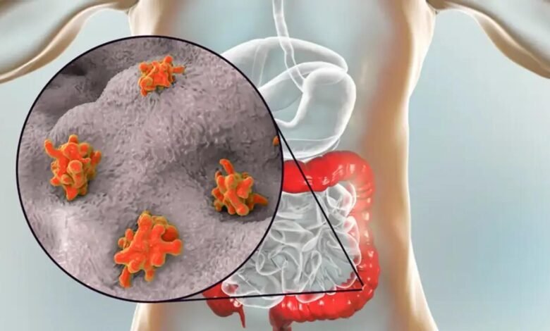
How Is Amebiasis Diagnosed by Healthcare Professionals?
Amebiasis is a parasitic infection caused by Entamoeba histolytica, a protozoan that primarily affects the intestines and, in some cases, the liver.

Amebiasis is a parasitic infection caused by Entamoeba histolytica, a protozoan that primarily affects the intestines and, in some cases, the liver. Healthcare professionals use a combination of clinical evaluation, laboratory testing, and imaging studies to diagnose amebiasis.
Prompt diagnosis is crucial for effective treatment, especially in severe cases such as amebic liver abscess. This article explores the diagnostic approaches for amebiasis, highlighting the role of nitazoxanide 500mg as a treatment option in managing the condition.
1. Clinical Evaluation
The diagnostic process often begins with a thorough clinical evaluation. Healthcare providers assess the patient’s symptoms, medical history, and potential risk factors for exposure to Entamoeba histolytica.
Symptoms of Amebiasis
The clinical presentation varies depending on whether the infection is intestinal or extraintestinal:
Intestinal Amebiasis
Common symptoms include diarrhea, abdominal pain, cramping, and sometimes dysentery (diarrhea with blood and mucus).
Extraintestinal Amebiasis
Symptoms of liver abscess include fever, right upper quadrant abdominal pain, weight loss, and hepatomegaly (enlarged liver).
Risk Factors
Certain factors raise suspicion for amebiasis, such as:
- Recent travel to endemic areas (e.g., tropical and subtropical regions).
- Poor sanitation or use of contaminated water.
- History of close contact with an infected individual.
2. Stool Examination
Stool analysis is a cornerstone of diagnosing intestinal amebiasis. It involves detecting Entamoeba histolytica cysts or trophozoites in fecal samples.
Microscopy
A wet mount or iodine-stained preparation of stool is examined under a microscope. The presence of cysts or trophozoites confirms the infection but does not differentiate E. histolytica from the nonpathogenic species Entamoeba dispar.
Antigen Detection
Enzyme-linked immunosorbent assay (ELISA) or immunochromatographic tests detect E. histolytica-specific antigens in stool samples. These tests are more sensitive and specific than microscopy.
Molecular Testing
Polymerase chain reaction (PCR) tests identify the DNA of E. histolytica in stool, providing definitive confirmation and differentiating it from other Entamoeba species.
3. Serological Testing
For extraintestinal amebiasis, particularly amebic liver abscess, serological tests play a significant role. These tests detect antibodies against E. histolytica in the blood.
Common Serological Methods
- Indirect Hemagglutination (IHA): Detects antibodies with high sensitivity.
- Enzyme-Linked Immunosorbent Assay (ELISA): Widely used due to its accuracy and rapid results.
- Positive antibody tests are particularly useful in non-endemic areas, where prior exposure is uncommon.
4. Imaging Studies
When extraintestinal complications like liver abscesses are suspected, imaging techniques are essential.
Ultrasound
A non-invasive and readily available method to detect abscesses in the liver. Typical findings include hypoechoic or anechoic lesions.
CT Scan or MRI
These advanced imaging modalities provide detailed visualization of abscess size, location, and surrounding tissue involvement. They are particularly useful in complicated cases or when surgical intervention is considered.
5. Colonoscopy or Sigmoidoscopy
In cases of severe intestinal amebiasis, endoscopic procedures like colonoscopy or sigmoidoscopy may be performed.
Findings
The presence of flask-shaped ulcers in the colon is highly suggestive of amebiasis. Biopsy samples can be collected for histopathological examination to confirm E. histolytica infection.
6. Differential Diagnosis
Amebiasis can mimic other gastrointestinal or hepatic conditions, so differential diagnosis is essential. Conditions to rule out include
- Bacterial dysentery (e.g., caused by Shigella or Salmonella).
- Inflammatory bowel disease (IBD).
- Giardiasis or other parasitic infections.
- Pyogenic liver abscess.
7. Role of Nitazoxanide 500 mg in Treatment
Nitazoxanide, an antiparasitic and antiviral agent, has emerged as an effective treatment for intestinal amebiasis, particularly for mild to moderate cases.
Mechanism of Action
Nitazoxanide inhibits the pyruvate:ferredoxin oxidoreductase enzyme essential for energy metabolism in protozoa. This disrupts the growth and replication of E. histolytica.
Dosage
The recommended dose of nitazoxanide 500mg for adults is 500 mg taken orally twice daily for three days. It is typically used as an alternative to first-line treatments such as metronidazole or tinidazole.
Advantages
Broad-spectrum efficacy against other intestinal parasites like Giardia lamblia and Cryptosporidium parvum. Favorable safety profile with minimal side effects.
Limitations
While effective for intestinal amebiasis, nitazoxanide is not suitable for treating extraintestinal forms like liver abscess. In such cases, nitroimidazole derivatives (e.g., metronidazole) remain the primary treatment.
8. Treatment of Extraintestinal Amebiasis
For amebic liver abscess and other severe manifestations, the treatment typically involves
- Metronidazole or Tinidazole: High efficacy against E. histolytica.
- Luminal Agents: To eradicate cysts in the intestines, drugs like paromomycin or iodoquinol are added after systemic treatment.
- Drainage of Abscesses: Rarely, percutaneous or surgical drainage may be needed for large or complicated abscesses.
9. Preventive Measures
Prevention plays a crucial role in reducing the incidence of amebiasis, particularly in endemic regions. Key measures include:
- Improved sanitation and access to clean drinking water.
- Proper hand hygiene and food preparation practices.
- Screening and treating asymptomatic carriers to prevent transmission.
Conclusion
Amebiasis diagnosis requires a comprehensive approach, integrating clinical evaluation with laboratory and imaging studies. Stool examination, serological tests, and imaging techniques are indispensable tools for identifying intestinal and extraintestinal infections.
Nitazoxanide 500 mg serves as a valuable option for treating intestinal amebiasis, offering a safe and effective alternative to traditional therapies. However, its role is limited in managing severe extraintestinal forms, where nitroimidazole derivatives remain the gold standard. Awareness of diagnostic methods and treatment options, coupled with preventive strategies, is essential to combat this parasitic disease effectively.


